
The KODAK 9000 3D Extraoral Imaging System
A versatile, cost-effective imaging solution for modern dental practices.
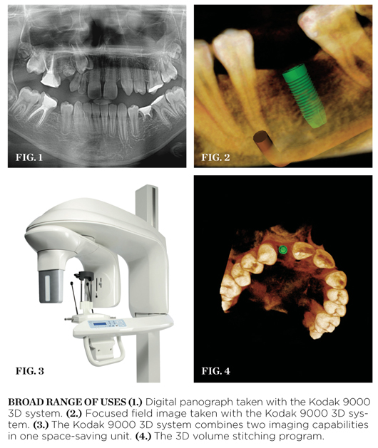
The Kodak 9000 3D system features both digital panoramic (Figure 1) and focused-field 3D (Figure 2) imaging modalities—two imaging systems in one attractive, space-saving footprint (Figure 3). It embodies an innovative and comprehensive approach by using complementary imaging modalities to enhance a user’s diagnostic abilities and to broaden the range of treatments a practice can offer. Evaluate bone quality and quantity, view periapical lesions, localize anatomic structures, pathologies, and more—the Kodak 9000 3D system is rapidly changing the way we view our patients and the way radiographic examinations are conducted.
3D patient studies are generated at an impressive slice thickness of 0.076 µm, the highest resolution in the industry. Precision digital detectors, combined with fiber-optic plate technology, optimize the signal-to-noise ratio and image quality. The added power of cone beam—a 50-mm x 38-mm focused field of view—helps the operator to visualize specific regions of interest with true anatomic accuracy.
As an added benefit, the Kodak 9000 3D system is now available with an optional 3D volume stitching program (Figure 4), enabling users to capture and visualize a larger view of a patient’s anatomy. This stitching capability is also available as an upgrade to units that predate its inception. The 3D stitching program combines up to three focused-field volumes, automatically constructing an extended-field image (as large as 85 mm x 66 mm x 37 mm). An outstanding solution for evaluating a variety of clinical case types, such as full-arch implant rehabilitation, multiple tooth impactions, pathology, and trauma, the 3D stitching program only adds to the Kodak 9000 3D system’s versatility. For those who need the option of cephalometric radiographs, there is also the Kodak 9000C 3D panoramic and cephalometric system.
Patient-Centered Approach
Because caring for patients’ health is a dental clinician’s top priority, the Kodak 9000 3D system was designed to adhere tightly to the ALARA principle, keeping radiation doses As Low As Reasonably Attainable without sacrificing image quality. The patient dose for 3D CBCT volumes is within the range of one to five periapical film images, markedly lower than larger-field CBCT systems. The Kodak 9000 3D system thus facilitates better patient care by providing more accurate and dependable treatment planning capabilities at a reduced patient dose.
Patient comfort is also a top priority, which is why the Kodak 9000 3D system features an open, face-to-face design to facilitate patient positioning and allows users to maintain eye contact and ease patient anxiety. For panoramic imaging, it is a true “one size fits all” solution: the system’s variable focal trough adapts to each patient’s anatomy. The system accommodates patients of all shapes and sizes and is easily accessible to patients in wheelchairs.
Intuitive Functionality
The Kodak 9000 3D system is not only convenient for patients—it’s convenient for doctors too. Simply select the desired imaging program, either 2D or 3D, and the system automatically positions the appropriate sensor. The intuitive, easy-to-learn imaging software is designed to have a short learning curve, requiring little training and allowing for smooth integration within a busy practice. With full 3D image reconstruction times of less than 2 minutes, the Kodak 9000 3D extraoral imaging system can reduce examination time and speed workflow. Rapid image acquisition and the ability to use multiple modalities in a single technology combine to boost a practice’s efficiency.
The 3D viewing software module includes a number of specialized image visualization tools and provides a variety of options for viewing and analyzing the image data to maximize the diagnostic yield and effectiveness of the images. With a built-in tool to “capture” key images and the capacity to burn 3D image volumes with a fully-functional 3D viewing module, the Kodak 9000 3D system is ideal for patient education and collaboration with other healthcare providers.
This article was written by David Gane, DDS. He has served as a consultant on dental imaging for a number of universities and corporations.
For more information, contact:
PracticeWorks Systems, LLC,
The Exclusive Maker of Kodak Dental Systems
Phone: 800-944-6365
Web: https://www.kodakdental.com
E-mail: info@practiceworks.com
Disclaimer
The preceding material was provided by the manufacturer. The statements and opinions contained therein are solely those of the manufacturer and not of the editors, publisher, or the Editorial Board of Inside Dentistry. The preceding is not a warranty, endorsement, or approval for the aforementioned products or services or their effectiveness, quality, or safety on the part of Inside Dentistry or AEGIS Communications. The publisher disclaims responsibility for any injury to persons or property resulting from any ideas or products referred to in the preceding material.


