
The Alphard—The Ultimate in CBCT
Belmont Equipment (Somerset, NJ) will introduce the cutting-edge, reliable technology of its Alphard 3D X-Ray Cone Beam CT scanner from Japan to the US market. The Quolis Series Alphard (Figure 1) provides multi-imaging from dental computed tomography (CT) to cephalo CT to meet the various diagnostic needs of small and large areas during a single exposure. The Alphard makes it possible to display and detect both hard and soft tissues, and to display profiles of faces in three dimensions (3-D) for uses ranging from implants to orthodontics.
Cone Beam Technology
Conventional CT scanners require separate scans of the mandible and maxilla, subjecting patients to unnecessary additional radiation. Using cone beam computed tomography (CBCT), scans of the mandible and maxilla are possible in just one exposure with the Alphard. CBCT technology directs a narrow cone beam of radiation for both parts of the jaw in just one digital image, providing a decrease in exposure and an increase in accurate images. The Alphard CBCT is accurate to 0.1 mm.
FLAT PANEL DETECTOR
Direct 3-D digital radiography is possible with the Alphard’s Flat Panel Detector (FPD) (Figure 2). By combining a cesium scintillator FPD with CT, three images are possible in one exposure. The FPD does not suffer the effects of the Earth’s magnetism, and thus provides sharp, clear, and distortion-free images. The FPD converts x-rays to light and then converts the light to electric charges using a photodiode and electric output. The dynamic range of expressive tones is 64 times 8 bits for reliable displays. Exceptional details within the image quality of broad areas are created by various voxel sizes. Voxels, which are more sophisticated than pixels, are 3-D cubes of specific sizes to create the ultra-fine details. Four separate modes provide the ability to vary voxel sizes for the type of image needed for diagnosis.
MODES
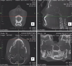
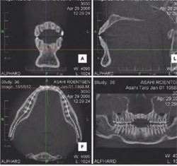
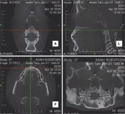
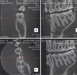
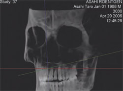
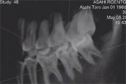
A built-in collimator automatically sets the exposure field according to exposure mode. Voxel sizes vary according to modes from 0.1 mm to 0.39 mm. Four modes are possible: C-Mode, I-Mode, P-Mode, and D-Mode. The C-Mode can cover a broad exposure area of 20 cm x 18 cm, which is exceptionally adequate for orthodontic images of almost the entire cranium, while differentiating between hard and soft tissues (Figure 3). I-Mode covers a broad exposure area of 10 cm x 10 cm, with accurate measurement of distance for implants (Figure 4). P-Mode provides 3-D images as well as panoramic images with a 15 cm x 15 cm exposure area (Figure 5). D-Mode provides a 5 cm x 5 cm high-resolution exposure area with accurate measurements of selected areas (Figure 6).
POSITIONING MECHANISM
Equipped with an adapted automatic positioning mechanism and an electrically driven chair, the Alphard moves automatically to the optimal exposure position by means of simply focusing the exposure-area indication beam on the patient. A remote control provides quick, accurate position operation.
NETWORKING AND SOFTWARE
The Alphard’s reception system and server are connected by local area network (LAN), providing operatories with the ability for one person to perform the x-ray from beginning to end. Patient data can be transferred to other facilities with ease by using various recording media.
Asahi Vision software (Asahi Roentgen Ind Co, Ltd, Kyoto, Japan) provides the ability to predict the position of a cuspid or any unerupted teeth, the thickness of the alveolar bone, and views of the molars, all parts of the head, and the mandible joint. A raysum image can also be created from the data received for the 3-D images, allowing multiple exposure types from a single exposure. Multiplanar reconstruction images at an arbitrary cross-section (tomographic images), 3-D image display, angle measurement, distance measurement, reversal black-and-white images, image density adjustment, and coloring are available. Asahi Vision software is also user-friendly, providing visible icons and patient-database control for editing and saving patient exposure conditions and modes. Necessary patient data and dates are included on each x-ray scan, creating an efficient and economic solution for file storage and retrieval.
ENDLESS DIAGNOSTICS POSSIBLITIES
The extremely large amount of information and high-accuracy 3-D images generated by the Alphard can expand diag-nostic capabilities. The geometric positional relations between cranial parts, such as an impacted tooth, temporomandibular joint, alveolar bone, cranial part, etc, can be accurately obtained. In addition, dentists can compare and evaluate pretreatment, posttreatment, preretention, and postretention 3-D images for more accurate planning and diagnostic treatment. The Alphard allows dentists to save time and money in lengthy explanations, image corrections, and model reconstructions for patient consultations. The Alphard’s variable-view 3-D images and raysum images provide distinct clarity for patients.
For more information or a brochure, contact:
Belmont Equipment
Division of Takara Belmont USA, Inc
Phone: 800-223-1192, ext. 1188
Web: www.belmontequip.com
E-mail: jsullivan@belmontequip.com
DISCLAIMER
The preceding material was provided by the manufacturer. The statements and opinions contained therein are solely those of the manufacturer and not of the editors, publisher, or the Editorial Board of Inside Dentistry. The preceding is not a warranty, endorsement, or approval for the aforementioned products or services or their effectiveness, quality, or safety on the part of Inside Dentistry or AEGIS Communications. The publisher disclaims responsibility for any injury to persons or property resulting from any ideas or products referred to in the preceding material.
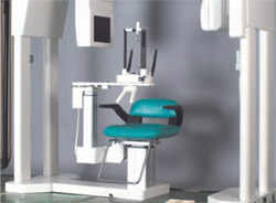 | 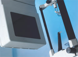 | |
| Figure 1 The Quolis Series Alphard CBCT. | Figure 2 The Alphard’s Flat Panel Detector (FPD). | |
 |  | |
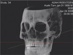 | 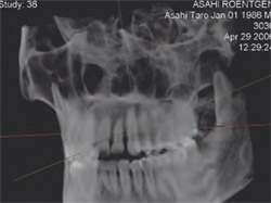 | |
| Figure 3 C-Mode provides a broad exposure area of 20 cm x 18 cm to distinguish hard and soft tissues, which is excellent for orthodontic treatment. | Figure 4 I-Mode provides a broad exposure area of 10 cm x 10 cm for sharp, clear images of selected areas, which is excellent for implant procedures. | |
 |  | |
 |  | |
| Figure 5 P-Mode provides panoramic or 3-D images of a broad exposure area of 15 cm x 15 cm. | Figure 6 D-Mode provides a high-resolution exposure area of 5 cm x 5 cm of selected areas for exceptionally accurate diagnosis. | |

