
An Alternative Treatment Modality for Transitionalizing a Removable Partial Denture to a Complete Denture
Bruce J. Goldman, DDS
During prosthetic treatment, patients often express concern that they may have to function without their removable prostheses while these appliances are being converted in a dental laboratory. The conversion may involve adding teeth or clasps to partials, relining removable partial dentures or complete dentures, or converting removable partial dentures to complete dentures. A common treatment dilemma involves the last scenario in which an interim appliance must be provided immediately after extracting teeth. Typically, in the case of a patient who must lose his or her remaining teeth in an arch, the dentist must withdraw a partial in an impression, send the impression to a commercial laboratory, and ask the laboratory to process new teeth to the existing partial. This process can take 1 to 2 days and requires the patient to relinquish his or her appliance for that time.
When the prosthesis is eventually returned to the patient, oftentimes the saddle will impinge upon the tissue because of the inherent inaccuracies of immediate denture placement. An alternative treatment modality is offered in this article, which provides a truly immediate conversion of a removable partial denture (RPD) to a complete denture.
CASE REPORT
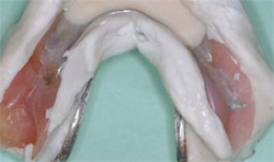
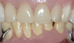
A 60-year-old woman presented with the chief complaints of her remaining lower teeth being loose and discomfort on chewing. Examination revealed that the patient used a maxillary complete denture and a mandibular RPD. Teeth Nos. 21 through 27 were present and Nos. 21 and 27 served as the abutments for the mandibular RPD. All of the remaining mandibular teeth had severe bone loss and extensive periodontal pocketing. Figure 1 shows the preoperative appearance of the remaining teeth. Note the purulence at the gingival margin on many of the teeth. Radiographically, virtually all of the teeth exhibited between 50% to 80% loss of vertical bone height.
The considered treatment options were:
- Periodontal treatment, selective extractions, and the fabrication of a new mandibular RPD.
- Extraction of all of the teeth except the canines, followed by surgical pocket elimination around the canines, endodontic therapy, and the fabrication of a mandibular overdenture using appropriate intraradicular retentive devices such as Zest® anchors (Zest Anchors, Inc, Escondido, CA), ERA® anchors (Sterngold Dental, LLC, Attleboro, MA), or Locator® abutments (Zest Anchors, Inc, Escondido, CA).
- Extraction of all of the remaining teeth, followed by implant placement and the fabrication of an implant-retained overdenture.
- Extraction of all of the remaining teeth, followed by the fabrication of a complete mandibular denture.
Regardless of the treatment modality chosen by the patient, she expressed a desire to never leave the office without mandibular teeth in place. The treatment options were discussed with the patient. Depending on how many teeth would be extracted we could either:
- Make a new transitional RPD to be delivered at the time of extraction.
- Modify her existing RPD by adding teeth Nos. 21 through 27. This could be accomplished by sending the appliance to a laboratory to extract teeth Nos. 21 through 27 on the model, approximate where the ridge would be after surgery, and add denture teeth and facial and lingual flanges to the RPD.
- Modify her existing RPD in-house at the same appointment as the surgical visit.
Because of limited financial resources, the patient elected to have all her remaining teeth extracted and the existing partial transitionalized in the office.
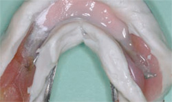
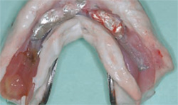
The patient was administered local anesthesia. Her mandibular partial was withdrawn within an alginate impression and set aside in a sealed bag (Figure 2). A flap was raised from teeth Nos. 21 through 27, and the teeth were extracted. An alveoloplasty was performed and the sockets were degranulated. The flap was closed with six 3-0 black silk sutures (Figure 3).
Dry foil (Jelenko, San Diego, CA) was cut to size and glued over the flap with iso-cyanoacrylate (Cyanodent, Ellman International, Hewlett, NY) (Figure 4). Jet Acrylic (Lang Dental Mfg Co, Inc, Wheeling, IL) in shade #66 was salt-and-peppered into teeth Nos. 21 through 27 of the impression holding the partial (Figure 5). Pink-fibered denture acrylic (Perm® Reline & Repair Resin, Coltène Whaledent, Inc, Cuyahoga Falls, OH) was then salt-and-peppered over the white acrylic (Figure 6). The impression was seated into the patient’s mouth and the acrylic was allowed to set to a doughy stage and then removed before the exothermic reaction of the acrylic occurred. The impression with the modified partial was then allowed to bench set in warm water until both acrylics were completely hardened (Figure 7).
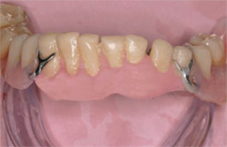
The dry foil, which adhered to the acrylic, was easily peeled off and the partial was trimmed and finished in our laboratory (Figure 8). The transitionalized denture was relined with a soft silicone material to provide more comfort for the patient (Figure 9). The final converted partial can be seen in place in Figure 10.
CONCLUSION
The patient wore this transitionalized denture through healing and through the fabrication phase of a new lower complete denture. This technique allowed us to inexpensively provide the patient with a comfortable transitional denture without ever having to give up the prosthesis for modification in a commercial laboratory.
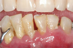 | 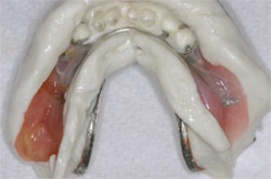 | |
| Figure 1 The preoperative appearance of the remaining teeth. Note the purulence at the gingival margin on many of the teeth. | Figure 2 The patient's mandibular partial was withdrawn within an alginate impression and set aside in a sealed bag. | |
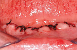 | 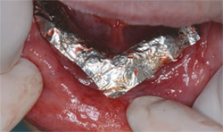 | |
| Figure 3 The flap was closed with six 3-0 black silk sutures. | Figure 4 Dry foil was cut to size and glued over the flap with alkyl 2-cyanoacrylate. | |
 |  | |
| Figure 5 Jet Acrylic in shade #66 was salt-andpeppered into teeth Nos. 21 through 27 of the impression holding the partial. | Figure 6 Pink-fibered denture acrylic was then salt-and-peppered over the white acrylic.The impres- sion with the embedded partial was reinserted into the patient's mouth so that the tissue side formed to the newly shaped mandibular ridge. | |
 |  | |
| Figure 7 The impression with the modified partial was allowed to bench set in warm water until both acrylics were completely hardened. | Figure 8 The dry foil, which adhered to the acrylic, was easily peeled off and the partial was trimmed and finished in the lab. | |
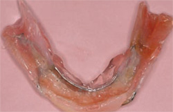 |  | |
| Figure 9 The transitionalized denture was relined with a soft silicone material to provide more comfort for the patient. | Figure 10 The final transitionalized RPD in place. | |
| About the Author | ||
 Bruce J. Goldman, DDS Bruce J. Goldman, DDS Private Practice Saugus, Massachusetts | ||

