
How to Liberate Dentists from Marginal Concerns
In restorative dentistry, marginal staining and discoloration, including gray, white, and brown lines, is a concern. The various types of discoloration have different generating mechanisms and causes, and general practitioners should treat each category differently.
White line—This is usually caused by a crack or fissure in the enamel. A white margin may also be a void at the interface between the bond and enamel. Enamel can be stressed from over-etching and from excess polymerization shrinkage of the restorative composite. One way to control the polymerization stress is by layering the composite. Another method for minimizing the possibility of a white line is to use a stronger bonding agent (such as those manufactured by Kuraray) and to avoid spaces at the interface between the bond material and the enamel.
Gray line—Typically this is caused by leaving too much bonding resin on the margin, allowing the background to be visible through the bonding layer. To prevent this, the bond should be spread by blowing air or wicked with a brush in order to maintain a thin film.
Brown line—This is the staining of the bond layer usually due to excess film thickness. A brown line can also result from microleakage at the interface between uncut enamel and the composite where the composite “laps” over the margins. First, the clinician must rule out caries; otherwise, the brown line can be eliminated by polishing.
Is phosphoric acid-etching or pre-etching necessary and effective for minimizing or eliminating marginal staining? Clinicians may need to apply phosphoric acid when using other self-etching agents, as there are many different types of self-etch products that do not perform at the same level as Kuraray’s self-etch products.
In their 10-year clinical evaluation, Akimoto et al1 concluded that most cases showed slight marginal changes during clinical function; however, these changes were not clinically significant. Their data demonstrated that placement of CLEARFIL™ Liner Bond 2 (Kuraray) self-etching adhesive system was acceptable for the clinical restoration of human teeth.
1 Akimoto N, Takamizu M, Momoi Y. 10-year clinical evaluation of a self-etching adhesive system. Oper Dent. 2007:32(1):3-10.
 |  |
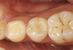 | 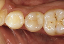 |
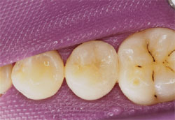
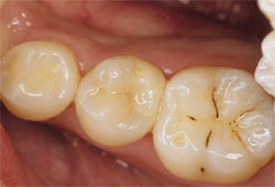
| Posterior case using CLEARFIL PROTECT BOND, CLEARFIL MAJESTY Flow, and CLEARFIL MAJESTY Posterior. Postoperative, 2-month, 1-year, and 2-year views.* (Dr. Naotake Akimoto, Tsurumi University, Japan) *No gaps, discolorations, or leakages in the margins. | |
For further information and to view the supporting figures and illustrations, please see the online interactive pdf, Technical Solutions for CLEARFIL Adhesives, at: https://kuraraydental.com/resources/01_technical_solutions_for_clearfil_adhesives.pdf.

