You must be signed in to read the rest of this article.
Registration on CDEWorld is free. You may also login to CDEWorld with your DentalAegis.com account.
We have entered a new era in dentistry in which technology and artistry have converged to provide the dental team with advanced digital tools and materials for replicating the intricacies of nature. The ability to precisely capture the tooth preparation using digital impression tools has eliminated the chairside limitations presented by the physical impression, such as tears, voids, and discrepancies. For the patient, the experience of impression-taking has become more comfortable. For laboratory technologists, receiving digital impression files electronically removes the inconsistencies inherent in creating a physical stone model and provides the precision information needed for creating a restoration that exhibits the unprecedented accuracy of fit, form, and function.
Much like the birth of implantology, CAD/CAM technology promises to be one of the major forces impacting the dental industry today and in the future. For the dental team, using advanced technologies in the practice and laboratory enables the merging of passion, experience, and dental knowledge with the precision of digital tools to create a new standard of patient care.
Change always brings with it the opportunity to move forward, and progress in not only educating oneself, but for the growth, and future of the practice and the laboratory’s business. Advances in technology provide a new workflow for the dental team that reduces turnaround times, streamlines fabrication, and provides patients with metal-free restorative solutions that exceed their expectations.
For the dental team, implementing digital impression scanning technology such as Planmeca’s PlanScanTM (www.planmecacadcam.com), PlanCADTM design, and PlanMillTM 40 milling has resulted in the dentist, dental assistant, and laboratory technician working as a close-knit team focused on providing the best practices in treatment. This interdisciplinary approach ensures the best possible outcome and experience for the patient.
Case Study
A female patient presented with a vertical fracture on tooth No. 9 (Figure 1). The clinician chose to extract the fractured tooth and place a BIOMET 3i implant (BIOMET 3i, www.biomet3i.com) with a screw-retained zirconia-milled abutment and restored the single central with a milled full-contour IPS e.max® crown (Ivoclar Vivadent, www.ivoclarvivadent.us), cut back for layering.
In one appointment, tooth No. 9 was extracted, the implant placed, and tooth provisionalized. Prior to provisionalization, an intraoral scan body was placed and digitally scanned. During the same appointment, the restorative B1 shade was determined and recorded using digital photography. The open-architecture STL electronic impression scan was transmitted to BIOMET 3i for model and abutment fabrication.
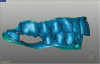
After receiving the model and milled implant abutment, the digital designer scanned the model using PlanScan CAD/CAM technology and imported the scan into PlanCAD design software. There, the anatomic morphology of the restoration was created, the optimal height of the contours was adjusted, the anatomy was matched to the adjacent and opposing dentition, and the proximal and occlusal contacts were designed (Figure 2). The CAD file was exported to the PlanMill 40 unit for milling. Figure 3 shows the full-contour restoration after milling and before crystallization and cutback.
For dental technicians, the single maxillary central incisor is perhaps the most challenging restoration to replicate. Color, translucency, internal characterization, surface texture, and luster all play a critical role in the success of matching the restoration to adjacent dentition.
The patient’s natural dentition appeared to have complex inner characterization, along with incisal translucency. The most challenging aspect of mimicking natural tooth No. 8 was in creating the same translucency of the incisal one-third. IPS e.max modifiers were used at the internal stain stage to achieve the correct incisal effect. The digital photos also were critical in maintaining the patient’s smile line. Closing the gingival embrasure between the patient’s two centrals was accomplished by adding to the mesial lingual interproximal aspects of the restoration.
Once the final restoration was completed, the clinician cemented the crown in place and had the patient schedule a follow-up appointment so the team could ensure gingival integrity. Figure 4 shows the final restoration in place a few weeks after cementation. The patient and team were pleased with the results.
Conclusion
As this case proves, dental practices and laboratories that embrace the latest in technologies can combine the precision of the machine age with custom artistry and solve challenging and complex cases. Excellent results can be achieved when technology, artistry, and nature converge.
Disclosure
This article was supplied by E4D Technologies.
About the Authors
Curtis E. Jansen, DDS
Curtis E. Jansen, DDS, completed his DDS and advanced education in prosthodontics at the University of Southern California (USC) School of Dentistry. He taught full time at USC and was director of implant dentistry in the Department of Restorative Dentistry. Dr. Jansen is a member of the American College of Prosthodontics, an active member of the Academy of Osseointegration, and a Fellow of the American College of Dentists. Currently, he has a full-time practice limited to prosthodontics and a dental laboratory in Monterey, California.
Frank S. Mercurio
Frank S. Mercurio has owned and operated Mercurio Dental Arts in Kensington, CA for more than 35 years. He received his dental technology degree from Diablo Valley College in Concord, California. He has been actively involved in implant restorative procedures since the early 1980s. For the past 6 years, Mr. Mercurio has been incorporating CAD/CAM procedures into his laboratory. In the past, he has presented live hands-on CAD/CAM courses involving stain glaze and add-on procedures to various groups.
Irma Perez, RDA, EF, CDD
Irma Perez, RDA, EF, CDD, is a registered dental assistant with extended function certification, a chairside digital designer, and a former removable technician. She has extensive Procera, SimPlant, and NobelGuide training. She has been using CAD/CAM for restorative dentistry with Curtis E. Jansen, DDS, for the last 8 years.