
New Burs and Strategies for Endodontic Access
Whenever a clinician faces endodontic access in the calcified tooth, a slower, and sometimes very different protocol should be followed.
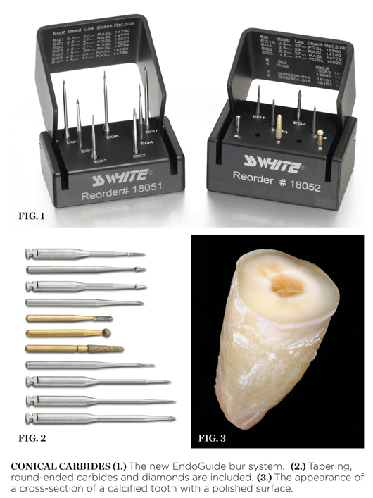
SS White Burs, Inc. has introduced the first conical carbides for endodontic access (Figure 1). These unique and minimally invasive shapes are based on the patented Fissurotomy® (SS White, Inc., www.sswhiteburs.com) cutting shape. These burs are part of a comprehensive endodontic access system (Figure 2). Included in this system are also tapering, round-ended carbides and diamonds. Because they are no longer state of the art when cutting endodontic access, round burs are missing from the system. Also missing are Gates Glidden burs because one or two EndoGuide burs can replace sizes 1 through 6 of the Gates Glidden burs for orifice enlargement.
Conical Carbide Burs
Many calcified teeth have visual clues that guide a clinician using advanced magnification safely into the tiny recessed pulp chamber. Polished or wet dentin is much like a polished rock. With magnification, subtle differences in color are enhanced and act like a bull’s-eye to guide the clinician. Figure 3 demonstrates the appearance of a cross-section of a calcified tooth with a polished surface (carbide bur).
For tactile dentistry, the conical carbide acts very differently than a round bur. For example, the EndoGuide’s tip size is half the tip size of the corresponding round bur. Equally important is the resultant shape cut with the conical carbide bur. When using a round bur for access, the resultant irregular and parallel-sided cavity walls work against the clinician when attempting to insert a hand file. When clinicians attempt to access the calcified tooth with a round bur, it is common for the bur to become slightly misdirected. When a file is inserted, it clunks into the bottom of the well. The clinician has no choice but to continue to tunnel deeper and go back and forth, inserting hand files into the fruitless bottom, and then burrowing deeper. In most cases, the wisp of pulp was higher up, like a trap door on the side wall.
When a machined, smooth, and conical-shaped access is cut with conical carbide, the visual contrast between dentin and pulp tissue remnants are tactilely accessible. Peri-cervical dentin is maintained and gouging is minimized.
Conical Carbide Film
Traditional access performed on a calcified tooth can be a very perilous journey. In contrast, when a conical shape is cut, the result can be far safer. The EndoGuide bur offers two unique advantages when taking a bur film. First, the tip is delicate enough to show precise tip location when compared to a round bur. The second advantage is the greater ease with which the radiograph can be taken without the bur slipping or falling out of the tooth during the capturing of the film. The conical carbide lightly binds to the dentin to provide stability while taking the film. Additionally, a small piece of border wax can be fastened simultaneously to the shank of the bur and tooth to provide temporary fixation for the taking of the film. The bur needs to be temporarily removed from the handpiece for this new type of check film.
Isolation
The commonly accepted practice in endodontics today is to place a single-hole rubber dam with the clamp on the tooth receiving the endodontic treatment. However, when accessing the calcified tooth, single-tooth isolation does not give the clinician a good three-dimensional (3-D) feel for root anatomy and angulation. The problem is compounded when there is a full crown present and the original anatomic landmarks are gone and replaced with something that can be very confusing. The clamp can also impede handpiece orientation as well as block radiograph passage if the clinician chooses to pause and take a radiograph for access location and direction verification. Endodontists often report that they remove the rubber dam entirely for difficult calcified cases to get a better feel for this 3-D procedure of canal discovery, and then replace the dam once the canal systems are safely discovered. The quadrant dam gives the best of both worlds.
This article was written by David Clark, DDS, who is a private practitioner in Tacoma, Washington.
For more information, contact:
SS White, Inc.
Phone: 800-535-2877
Web: www.sswhiteburs.com
Disclaimer
The preceding material was provided by the manufacturer. The statements and opinions contained therein are solely those of the manufacturer and not of the editors, publisher, or the Editorial Board of Inside Dentistry.


