
The Spectra System
Caries visualization is improved using fluorescent technology.
Early detection is often the key to effective caries management. Although the tooth may appear healthy on the surface, its real condition underneath —especially in the case of fissure caries—is often hard to detect. Using an explorer can lead to cavitation of early lesions, preventing remineralization, or may lead to an acceleration of the caries process when the weak enamel overlaying the demineralized area is physically broached by an instrument.
Enhanced caries detection, which supports minimally invasive treatment regimens, makes Spectra an innovative, indispensable tool. Spectra offers reliable tooth-by-tooth detection of fissure caries and caries on smooth surfaces. While other devices use numeric indicators to signal the presence of decay, Spectra uses a color system with software analysis to provide a picture of the tooth with visual representation of where tooth structure is deteriorating. Spectra’s LEDs project high-energy blue light onto the tooth surface, the wavelength of which stimulates bacteria to fluoresce red and healthy enamel to fluoresce green.
Spectra is similar to an intraoral camera in appearance and function. Yet, where an intraoral camera has white LEDs surrounding the lens, Spectra has an array of six LEDs emitting a 405-nm blue-violet light. Spectra connects to the operatory computer via a USB connector and is run by proprietary software. The device is sheathed in a single-use disposable intraoral camera sleeve for sterility and an autoclavable rubber “spacer” is placed over the sheath at the end of the lens. The spacer eliminates ambient light and maintains a consistent distance between the device and the tooth surface, so images are reproducible and consistent. Spectra is both self-calibrating and portable, making it easy to use in offices with multiple operatories.
With its instantaneous software analysis, the captured image of the tooth shows enamel breakdown resulting from caries in a graphic representation similar to weather radar images. Images can be saved to monitor areas or to gauge the effectiveness of remineralizing therapy.
It has long been recognized that Streptococcus mutansproduces special metabolites called porphyrins, which fluoresce when exposed to 405-nm light. This fluorescence is detected by the Spectra device and visually identified. The denser the bacterial colonization, the more intense the red fluorescent signal will be. The software analysis then provides an estimate of the depth and extent of the caries based on the color map produced, depending on the degree of fluorescence. The image and corresponding color map also help to assist the patient in understanding why the treatment plan being suggested is recommended despite a possible absence of any tooth sensitivity.
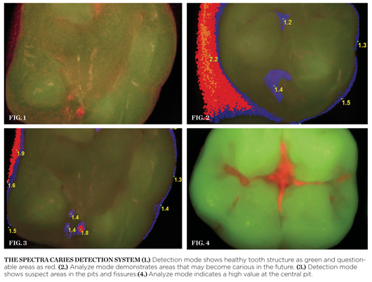
A healthy tooth may demonstrate slight fluorescence in the pits and fissures when observed under detection mode, but when switched to analyze mode, values should be 0 in these areas. A tooth with a similar appearance under detection mode (Figure 1) may, when switched to analyze mode, demonstrate some values in the pits and fissures (Figure 2). Low values in the pits and fissures may indicate continued observation of these areas or suggest that either remineralization procedures be used or sealants be placed. Teeth that show more extensive red fluorescence in the pits and fissures (Figure 3) may demonstrate higher values in these areas (Figure 4), leading the practitioner to perform conservative restorations rather than just observation until the caries have penetrated deeper and wider.
For more information, contact:
Air Techniques
Phone: 800-AIR-TECH
Web: https://www.airtechniques.com
Disclaimer
The statements and opinions contained therein are solely those of the author and not of the editors, publisher, or the Editorial Board of Inside Dentistry. The preceding is not a warranty, endorsement, or approval for the aforementioned products or services or their effectiveness, quality, or safety on the part of Inside Dentistry or AEGIS Communications. The publisher disclaims responsibility for any injury to persons or property resulting from any ideas or products referred to in the preceding material.
About the Author
This article was written by Gregori Kurtzman, DDS.


