
Attachment Adaptation to an Existing Maxillary Overdenture
Allen L. Schneider, DDS; and William A. Lobel, DMD
Because of the increased need for maintenance of existing removable prostheses—whether complete, partial, or overdenture—both the clinician and the patient have many challenges to face. Demographic studies have shown that the dental profession must be prepared to meet the prosthetic needs of an ever-increasing, longer-living patient population.1
When proper diagnosis, treatment planning, and, most importantly, patient compliance are achieved, the overdenture has been a proven mainstay of conservative prosthodontic treatment.2 The many advantages of root retention include: alveolar bone maintenance, better prosthesis support, proprioceptive feedback, esthetics, and psychological benefits.
This article demonstrates a technique to salvage an existing maxillary overdenture. The patient presented with carious root structure around the amalgam overdenture abutments on the second bicuspid and two canines. The residual maxillary ridge mucosa was healthy. The preexisting denture fit well and the esthetics were pleasing to the patient. The patient expressed a strong desire to keep his roots and avoid surgery.
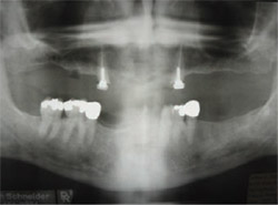
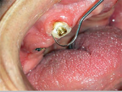
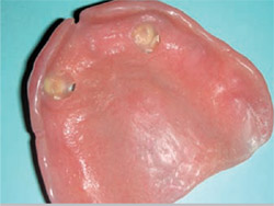
Decay was removed from the cuspid roots, exposing enough sound structure to proceed with treatment (Figure 1 and Figure 2). The second bicuspid was not restorable and subsequently extracted. The panoramic radiograph demonstrated previous successful endodontic therapy and sufficient root structure in the bone (Figure 3).
Locator attachments (Zest Anchors, Inc, Econdido, CA) were chosen to treat the cuspids to provide retention and stability in addition to restoring the previous support function of the remaining roots. Locators can be used to attach to roots with or without a coping in a direct or indirect technique. This case will demonstrate their use in a direct technique without a coping.
ROOT PREPARATION
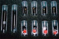
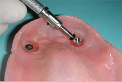
The next step was to prepare the root to receive the Locator post. The Locator Root Attachment Kit (Zest Anchors, Inc) (Figure 4) contains a sized latch-type post bur to prepare the canal space (Figure 5). At least 5 mm of gutta-percha should be left as an apical seal. The post may be shortened if necessary. The spot-faced bur was then used to create an internal dentinal seat for the Locator female post (Figure 6). If needed, 10°- or 20°-angle correction posts may be used to correct divergent roots. It is recommended by the manufacturer to parallel the path of insertion of the Locator attachments for root form prosthesis to less than 10° of divergence. Angle corrections for root form attachment of Locator-retained prosthetic appliances can be easily accomplished using the various angles of the female Locator post. Nylon components can be quickly and easily removed using the Locator Tool if a change in retention strength is necessary.
PREPARATIONFOR CEMENTATION
Before post cementation, Metal Primer II (GC America Inc, Alsip, IL) was applied to the serrated metal post portion to enhance bond strength to the dual-cure G-Cem™ resin cement (GC America, Inc) (Figure 7). The cement was placed in the clean, dry canal and the posts were seated using the black plastic alignment rods (Figure 8). The cement was light-cured, thus allowing quick removal of coronal excess (Figure 9).
DIRECT PICK-UP
The metal housings containing the male nylon processing components were then placed onto the posts cemented into the cuspid roots. The intaglio surface of the denture was hollowed out in the cuspid areas to allow complete intraoral seating of the denture without interference from the bulk of the two housings. A small relief hole was made in the denture to allow for extrusion of excess resin (Figure 10).
The superior portions of the metal housings were microetched and treated with the same metal primer to increase the bond strength of the metal housing to the luting resin (Figure 11). The housings, with included white spacer rings, were returned to the Locator females. A direct pick-up of the housings in the denture was accomplished using pink Triad® Gel (DENTSPLY International, York, PA) as the luting agent. A thin layer of Triad Bonding Agent (DENTSPLY International) was applied to the acrylic of the denture in the hollowed out area and light cured for 10 seconds to ensure a strong bond between the composite Triad gel luting agent and the denture acrylic. The resin was light-cured through the denture intraorally and the process was repeated for the other attachment (Figure 12 and Figure 13).
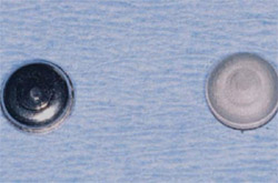
After the denture was adjusted and polished, the occlusion was verified (Figure 14). The Locator Tool was then used to remove both nylon processing components from the metal housings incorporated in the denture (Figure 15). Retentive nylon male components of various color-coded retentions can then be placed as dictated by the needs of the patient. The lightest-retention blue Locator retentive male, at 1.5 lbs, is generally recommended at delivery (Figure 16).
DISCUSSION
After the rehabilitation of the maxillary canines, the patient was instructed in the proper care of his root attachments by using a daily fluoride gel and performing meticulous plaque control. Regular recall appointments were scheduled to check for patient compliance and appropriate tissue fit of the denture. Relines will be performed as needed to ensure proper functioning and longevity of the attachments in addition to promoting long-term health of the supporting mucosa. The patient was extremely satisfied to have avoided extraction of his remaining cuspid roots and the ability to maintain his current denture. He was also pleased to gain the extra retention and stability from this treatment modality.
CONCLUSION
By respecting the patient’s wish for a conservative treatment and using Locator attachments to salvage an existing maxillary overdenture, the author was able to satisfy the patient and promote a healthy oral environment.
References
1. Douglass CW, Shih A, Ostry L. Will there be a need for complete dentures in the United States in 2020? J Prosthet Dent. 2002;87(1) 5-8.2. Stanwich LJ. The Tooth and Mucosa Supported Overlay Denture. In: Clark J. Clinical Dentistry. New York, NY: Harper & Row Publishers; 1976:1-12.
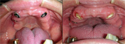 |  | |
| Figure 1 and Figure 2 Decay was removed from the cuspid roots, exposing enough sound structure to proceed with treatment. | ||
 | ||
| Figure 3 The panoramic radiograph demonstrated previous successful endodontic therapy and sufficient root structure in the bone. | ||
| Figure 4 The Locator Root Attachment Kit. | ||
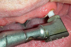 | 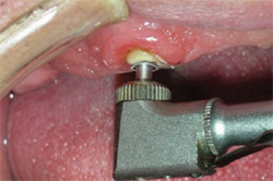 | |
| Figure 5 A sized latch-type post bur is used to prepare the canal space. | Figure 6 The spot-faced bur was used to create an internal dentinal seat for the Locator female post. | |
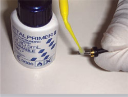 | 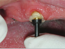 | |
| Figure 7 Metal Primer II was applied to the serrated metal post portion to enhance bond strength to the dual-cure G-Cem resin cement. | Figure 8 The posts were seated using the black plastic alignment rods. | |
 |  | |
| Figure 9 The cement was light-cured, allowing quick removal of coronal excess. | Figure 10 A small relief hole was made in the denture to allow for extrusion of excess resin. | |
 |  | |
| Figure 11 The superior portions of the metal housings were microetched and treated with the same metal primer. | Figure 12 and Figure 13 The resin was light-cured through the denture intraorally and the process was repeated for the other attachment. | |
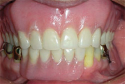 |  | |
| Figure 14 After the denture was adjusted and polished, the occlusion was verified. | Figure 15 The Locator Tool was used to remove both nylon processing components from the metal housings incorporated in the denture. | |
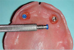 | ||
| Figure 16 The lightest-retention blue Locator retentive male, at 1.5 lbs, is generally recommended at delivery. | ||
| About the Authors | ||
 Allen L. Schneider, DDS Allen L. Schneider, DDS Private Practice Springfield, Virginia | ||
 William A. Lobel, DMD William A. Lobel, DMD Clinical Assistant Professor Department of Prosthodontics and Operative Dentistry Tufts University School of Dental Medicine Boston, Massachusetts Private Practice Saugus, Massachusetts | ||

