
Do What They Want So You Can Give Them What They Need
Chris Stevens, DDS
A 17-year-old adolescent boy presented to the dental office. Both he and his mother were present and indicated that they were not happy with his smile. He related that he had a peg tooth on one side and a missing tooth on the other. He wanted his smile enhanced by increasing the size of the peg tooth in time for senior pictures, which were scheduled in 3 weeks. Once his senior pictures were taken, they wanted to replace the missing tooth.
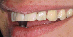
On examination, it was determined the patient was congenitally missing the right lateral incisor and had a peg left lateral incisor. The missing lateral was currently replaced by his orthodontic retainer. Incisal chipping of the right and left central incisor, a small diastema between the central incisors, and a reverse smile curve were also noted (Figure 1; Figure 2; Figure 3).
The patient’s mother had heard of implants from the orthodontist and felt that was the way she wanted to proceed with the right lateral incisor. For senior pictures she wanted the left lateral incisor bonded.
Treatment Options
Potential treatment options were discussed. The first option was to perform direct resin bonding on the left lateral incisor for senior pictures as the patient’s mother requested. Once the pictures were taken, implant procedures could be started on the right lateral incisor. The main concern with this option was that although it would satisfy the smile enhancement desired for pictures, restoring the lateral incisors would not correct the chipping and diastema in the central incisors or correct the reverse smile curve. Also, after further discussion, the patient expressed a desire for whiter teeth; therefore, performing individual tooth care on his smile could result in inconsistent color.
The second option consisted of an implant-supported porcelain crown for the right lateral incisor in addition to three porcelain veneers on the remaining incisors. This option would correct all of the issues, including the reverse curve and inconsistent color. The primary concern for this option was lack of time.
The author’s preference was the second option. It would be the best long-term option and it would produce a highly esthetic, controlled result. However, to have the second option considered by the patient’s mother, an interim treatment plan was needed that would satisfy her concern for the upcoming pictures.
It was decided that a laboratory-fabricated interim resin-based crown could be used to correct the size deficiency of the peg lateral incisor. This procedure would be easier than direct bonding of the peg lateral and would still produce a very satisfactory result. In addition, direct bonding could be performed on the central incisors to repair the incisal chipping. This would satisfy the patient and his mother for senior pictures as well as allow the best treatment plan available to the patient, but at a later date when time constraints were removed.
Treatment
After acceptance of the second option by the patient and his mother, maxillary and mandibular impressions were taken along with a centric occlusion bite registration and color mapping. These items were sent to the laboratory for fabrication of the resin-based interim crown. This crown was to be placed over the unprepared peg lateral incisor. Therefore, instructions to not prepare the tooth on the cast were given to the laboratory (Figure 4).
At a subsequent appointment, direct bonding was performed on the two central incisors. Recommended protocols for etching and adhesive placement were completed. A dentin-shaded hybrid resin was placed followed by incisal-shaded microfilled resin. Recommended shaping and polishing techniques were also performed.
Once the central incisors were completed, the left lateral incisor was treated. Light roughening of the enamel surface was completed with a fine diamond bur. Again, recommended protocols for etching and adhesive placement were completed. The resin interim crown was etched and silanated. The adhesive was placed and cured for the recommended time.
A dual-cure resin cement was placed in the crown and the crown was fully seated. Without dislodging the crown, excess material was removed with a rubber-tipped instrument. The resin cement was then cured according to the manufacturer’s recommendations. Final cement removal was performed with 12-fluted carbide burs and polishing cups.
Slight adjustments to the shape were made and the occlusion was adjusted as needed. The end result was very satisfactory to both the patient and his mother (Figure 5 and Figure 6).
CONCLUSION
Patients may often present with an idea of what they want, especially in this day of easy access to information on procedures. The mother of this young man knew of available procedures regarding his lateral incisors. But convincing her to say, "Yes," to more comprehensive care required an alternative that would satisfy the main reason for their presentation to the office (ie, the upcoming senior pictures). Once they were assured that what they were asking for could be completed, they were very receptive to accepting to more comprehensive care.
Acknowledgment
The author wishes to thank Arrowhead Dental Laboratory, Sandy, Utah, for fabrication of the interim crown, and to acknowledge that he has received honoraria from Arrowhead Dental Laboratory.
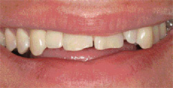 | 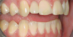 | |
| Figure 1 The unretracted frontal view. Note the reverse smile curve and incisal chipping. | Figure 2 The retracted right lateral view. Note the replacement of the congenitally missing right lateral incisor with the orthodontic retainer. | |
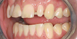 | 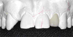 | |
| Figure 3 The retracted left lateral view. Note the peg lateral and the visibility of the orthodontic retainer, which is evident while smiling. | Figure 4 The laboratory-processed interim crown. | |
 | 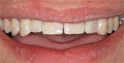 | |
| Figure 5 The unretracted left lateral view. The interim crown is seated. The mesial emergence profile will be enhanced in the final restoration. | Figure 6 The unretracted frontal view. Note the more level incisal plane of the treated teeth as the patient requested. Implant restoration of the right lateral incisor could now be started. | |
| About the Author | ||
 Chris Stevens, DDS Chris Stevens, DDSPrivate Practice Sun Prairie, Wisconsin | ||

