3D Imaging and Cone Beam CT is “Where the Action is!”
Scott D. Ganz
For as long as clinicians have been placing and restoring modern dental implants, there have been periapical and panoramic imaging films to help guide the process. While the gold standard for years, these two-dimensional (2-D) film images are limited by their inherent distortion factors, providing little information regarding bone density, and the indisputable non-interactive nature of the film itself. Digital radiography, while a major step in the right direction, is still 2-D, although it does allow for some interaction to enhance diagnostic interpretation. When computed tomography (CT) scans became available for dental applications, to improve the understanding of three-dimensional (3-D) properties of the jaw bones, patients were directed to medical and/or radiological facilities where CT scans could be taken. The original films from these scans gave clinicians a lot of information but no interactive ability, and were cumbersome because of their large size. With innovative software applications, like SimPlant (CSI-Materialise, Glen Burnie, MD), a new world of anatomical realization was unveiled, allowing for true, undistorted representations of the mandible or maxilla, combined with an array of tools for accurate treatment planning of dental implants.
Yet most of the world was still not ready for prime-time CT scanning of their patients, even though there was a growing awareness of the importance of the information gained from these advanced imaging technologies. It was not until the introduction of smaller, in-office cone-beam CT (CBCT) scanning machines in the late 1990s that another world of opportunity became available. Today, there are many different types of CBCT machines on the market, with more on the way. These machines are small enough to fit in almost any dental office, allowing for instant access to the technology. Just as the panoramic radiograph revolutionized the industry when it arrived on the scene, CBCT is now having great impact on everyday clinical practice. The introduction of these machines has been a huge catalyst to focus the dental industry, especially in the areas of implants and simple to complex oral surgery procedures. CBCT takes presurgical planning to the next level by leaps and bounds.
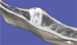
CBCT scans allow for undistorted, 3-D assessment of the maxillofacial complex. While conventional CT imaging usually involved one single arch on film, the entire skull can now be visualized for a better understanding of the actual maxillo-mandibular relationships (Figure 1). The advent of 3-D reconstruction using CT now empowers the clinician with simulating the plan for implant placement, bone grafts, or orthognathic surgical procedures in a true and accurate virtual environment. There are three basic views that a CT scan will provide: panoramic, axial, and cross-sectional. The fourth view contains the 3-D reconstruction, and may differ in quality depending on the scanning machine and the software that is used to manage the CT data. The ability to visualize the maxillary and mandibular alveolar complex in cross-section affords the clinician with a wealth of information (Figure 2). The teeth are in centric occlusion, yielding a view that can be quite revealing regarding the true trajectories of the roots and limitations of surrounding bone.
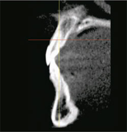
The standard CBCT views are particularly advantageous when planning for implant placement. Figure 3A illustrates a cross-sectional NewTom 3G CBCT (Dent-X®, Elmsford, NY) image of a hopeless tooth and surrounding alveolus in the anterior mandible. A one-piece implant (AdVent, Zimmer Dental Inc, Carlsbad, CA) was then simulated using a separate interactive software application (SimPlant) (Figure 3B). The resulting plan is a true schematic representation of the implant to be placed, allowing the clinician to accurately quantify and then manage the receptor site for the desired reconstruction. Further enhancements now allow for the CBCT images to be evaluated in 3-D, with computer-assisted design (CAD) representations of the actual implant(s) to be used (Figure 3C). Depending on the software, there are libraries of all of the most common implant systems to help in the planning process. The ability to rotate and position the implant with the abutment projection (yellow) in all four views defines true 3-D interactivity to achieve the most accurate placement of the implant(s). The capability of accurately assessing the bone for implant placement is no longer limited to fully edentulous cases. If the CBCT machine is readily available in the office, it becomes a routine tool that takes the guesswork even out of a case that involves only one or two implants in the esthetic zone, or above the inferior alveolar nerve in the posterior mandible.
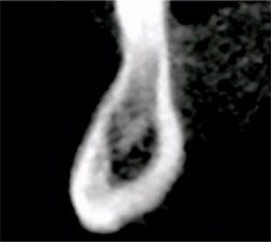
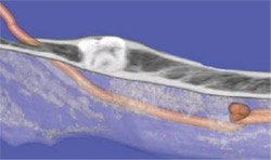
The role of CBCT in routine oral surgery procedures cannot be overemphasized. When evaluating a potential hor-izontally impacted molar, the axial view can be quite revealing. Figure 4 illustrates a molar that is not only horizontally impacted, but rotated to the lingual as well (see arrows). There was decay noted on the occlusal surface of the molar that was causing the patient pain and discomfort. The cross-sectional view of the posterior mandible revealed the lingual orientation of the impacted tooth, as well as its relationship to the underlying inferior alveolar nerve (Figure 5, seen in orange). This vital information can help clinicians understand the 3-D relationship of important anatomy, including bone, soft tissue, blood vessels, and nerves. The information can be further explored when the 3-D reconstruction can be sectioned in an axial plane to clearly reveal the true position of the tooth (Figure 6A). Based on this information, the correct treatment option and surgical approach can be determined. Having the opportunity to trace out the path of the mandibular nerve, while changing the bone density to allow for a transparent view, can deliver views of the undistorted anatomical relationships that have never been seen before (Figure 6B).
CONCLUSION
CBCT technology is here to stay. The intent of this brief article is to introduce some of the uses for this evolving technology. CBCT has empowered clinicians all over the world with advanced capabilities that will allow for better diagnosis, more accurate treatment planning, and improved outcomes for our patients. With the vastly improved ability to assess vital anatomical structures, significantly reduced radiation over conventional CT, ease of use, compatibility with most third-party software/hardware applications, and determination of bone density, the tools for presurgical planning have now been taken to a higher level. We need not be bound by two-dimensional concepts in a three-dimensional world.
DISCLOSURE
The author is a consultant for Materialise and Zimmer Dental.
For More Information
Dent-X
Phone: 914-592-6100
Web: www.dent-x.com
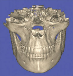 |  | |||
| Figure 1 Full skull visualized from a CBCT scan. | Figure 2 Cross-sectional view reveals both maxilla and mandible arches in occlusion. | |||
 |  | |||
| Figure 3A A cross-sectional image identified as a potential implant receptor site. | Figure 3B A simulated realistic schematic implant can be positioned to maximize success. | |||
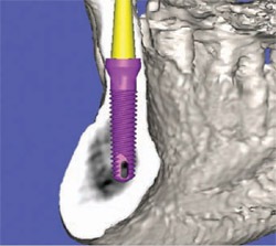 | 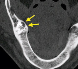 | |||
| Figure 3C Further enhancement of the 3-D reconstruction can be visualized within SimPlant from the NewTom 3G CBCT dataset. | Figure 4 When evaluating horizontally impacted teeth, the axial view can be quite revealing. | |||
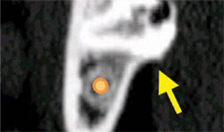 |  | |||
| Figure 5 The relationship between the tooth, the bone, and the mandibular nerve (orange) can be visualized in this cross-sectional view. | Figure 6A Using advanced tools to section the 3-D reconstruction aids in the planning of surgical extraction. | |||
 | ||||
| Figure 6B Adding transparency to the 3-D reconstruction aids in detecting the tracing of the inferior alveolar nerve, shown in orange. | ||||
| About the Author | ||||
| ||||