
Computed Tomography Imaging Is Changing Implant Dentistry
Cone beam volumetric imaging is relatively new to the dental industry. In 2005, the number of cone beam scanners on the market significantly increased. The spiral motion image capture used in cone beam tomography is much faster than that of traditional computerized tomography (CT) and, importantly, this type of imaging exposes the patient to approximately one-tenth of the radiation used in traditional CT scanners. In dentistry, cone beam scanners are being designed with space and patient comfort in mind. For example, upright seating is used in cone beam scanners with the x-ray tube and panel detector rotating around the patient’s head.
Cone Beam CT imaging software assists clinicians in visualizing a patient’s anatomy in a clinically meaningful way. Cone beam CT imaging is the preferred radiological modality for dental implantologists because of the high quality of the images, software capabilities, ability to manipulate and use information for surgical planning, and lower doses of radiation than traditional CT.
CT imaging in dentistry is evolving rapidly. Enhancements have created a new classification of CT scanner, marketed as ILUMA Flash CT® Scanner (IMTEC Imaging, Ardmore, OK) (Figure 1). Flash CT is an advanced program resulting in sharper CT images and reduced radiation exposure. Flash CT imaging allows clinicians to illustrate the recommended implant treatment plan to patients. In a statement, Ronald A. Bulard, DDS, a director of IMTEC Imaging, stated, “The added technology of the ILUMA scanner impresses my patients and contributes to my success of selling optional, out-of-pocket implant treatment plans.” Dr. Bulard attributes his ILUMA scanner to a 25% increase of patient treatment acceptance at his Dental Implant Centres in Ardmore, Oklahoma, and New York City.
Access to a Flash CT scanner significantly simplifies the process of evaluating treatment planning and achieving successful implant acceptance. Because the scanned images are available immediately, the patient does not lose interest or talk themselves out of implant treatments, which are often classified as elective by health insurance companies.
Flash CT imaging provides a necessary perspective in a variety of cases requiring an advanced amount of information. Examples of other applications of Flash CT in diagnosis and treatment planning are periodontal problems, impacted teeth, endodontic issues, temporomandibular joint conditions, jaw pathology, and airway analysis. Flash CT images from IMTEC Imaging provide important information about nerves, soft tissue, and bone, thereby assisting clinicians in designing successful treatment strategies. An added advantage of using Flash CT images when diagnosing patients and evaluating treatment options is the clinician’s ability to visualize the patient’s anatomy in 3-dimensional versus flat, 2-dimensional images. Three-dimensional volumetric software simplifies the assessment of anatomic considerations and prosthetic goals for implant surgical planning. In addition, 3-dimensional software accurately determines bone dimensions and quality through density shading. This advanced software determines information about the axis orientation of the alveolar bone for successful implant alignment. It identifies common internal anatomy needed to evaluate implant placement including jaw boundaries, adjacent teeth, nasal fossa, mandibular canal, maxillary sinus, mental foramen, and incisive canal. It also detects pathology to be avoided for implant success. Three-dimensional software is also capable of shading images to differentiate varying densities of facial structures. Grayscale shading results in the ability to view the relationship of common internal anatomy. Some versions of volumetric software, such as the software used in the ILUMA scanner, also permit clinicians to color code an image by density, further distinguishing anatomical structures. Color coding anatomical features enables the clinician to view the relationship of features while planning implant cases, such as nerves and nasal cavities in relation to the density and length of the mandible or maxilla.
Image data may be obtained for a complete dental/maxillofacial volume or a limited region of interest. With the current generation of cone beam scanners, scan times for these types of images vary from approximately 40 seconds to 1 minute for the complete volume and as little as 10 seconds for a regional scan. The next generation ultra cone beam scanners (such as IMTEC Imaging’s ILUMA) can take full-volume scans in 40 seconds or less.
Amorphous silicone flat-panel detectors, which are found in ILUMA Flash CT scanners, provide orthographic images (Figure 2). These images are sharp and dimensionally accurate, which is vitally important to treatment planning, especially in implant surgery cases. The dimensions of sensors vary by machine, but ILUMA offers 2 sizes of displays for dental and oral maxillofacial uses.
Volumetric software programs also enable a clinician to segment images to help evaluate the specific region of interest. Segmentation literally cuts the volume rendering, conceding top views, side views, and CT slices. CT slices are extremely thin, usually ranging from 0.3 mm to 0.1 mm.
Some software programs, including the one found in ILUMA, allow dentists to transform the digital images into STL (stereolithography format, or ASCII or binary file). ILUMA V-Implant software provides features allowing the exportation of STL files to 3-dimensional printers, thereby allowing modeling and fabrication of surgical guide stents. Imaging stents may be placed to show exact angulations and placement of a proposed implant. Using STL files to create a model helps the clinician see the exact angulations to use when placing the implant to ensure pleasing esthetics and security of the prosthetic tooth or teeth.
Cone beam CT imaging is quickly becoming the standard of care in implantology; however, the technology is evolutionary in nature. The software and hardware in CT technology is continually improving, making the cost of purchasing an ultra cone beam scanner a deterrent in private dental practices. The current generation of cone beam scanners is generally available only in imaging centers, hospitals, or group practices. The cost of the machine reduces patient and clinician access to this advanced technology and costs of imaging examinations are usually greater than that of film. In an additional conversation, Dr. Bulard noted that in his view if clinicians could obtain access to cone beam CT technology without dedicating a great amount of business capital into a depreciating technology, the use of cone beam scanners would rise exponentially.
CONCLUSION
Ultra cone beam tomography is quickly increasing the quality and accuracy of radiographic dental care. Used in a wide variety of cases requiring additional information for treatment planning and for detection of possible anatomical problems, cone beam CT scanners are literally changing the entire field of dentistry.
Implant dentistry will benefit greatly from Flash CT technology. Displaying a patient’s anatomy in a meaningful way, ultra cone beam scanners allow implantologists to plan cases thoroughly, obtain accurate measurements, manipulate data, and create surgical stents and models. These features save time in the planning and surgical stages of implant cases.
For More Information
IMTEC Imaging, LLC
Phone: 1-866-458-6228
Web: www.ilumact.com/
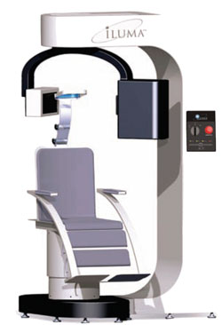 | 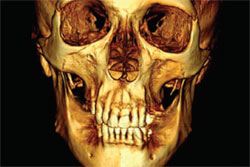 | |
| Figure 1 The ILUMA Flash CT scanner. | Figure 2 Silicone flat-panel detectors found in ILUMA Flash CT scanners provide orthographic images. |

